New Research: Cortical Layer 3 Pyramidal Neurons and Visual Spatial Working Memory

Visual spatial working memory, a core cognitive process, is dependent upon a cortical network composed of multiple nodes, including primary visual, posterior parietal, and dorsolateral prefrontal cortices. Within this network, information essential for proper visual spatial working memory function is transferred via projections furnished by pyramidal neurons located primarily in cortical layer 3.
Across the primate cortical visual spatial working memory network, layer 3 pyramidal neurons exhibit numerous morphological and functional differences. These differences are due, at least in part, to regional differences in the transcriptomes of these cells.
To improve our understanding of the transcriptomes of layer 3 pyramidal neurons from three nodes of the human visual spatial working memory network, Pitt Psychiatry investigators John Enwright, PhD, Dominique Arion, PhD, and David Lewis, MD, in collaboration with colleagues from the Rangos Research and UPMC Genome Centers, applied RNA sequencing to pools of layer 3 pyramidal neurons, collected individually by laser micro-dissection, from primary visual, posterior parietal, and dorsolateral prefrontal cortices of 39 postmortem subjects without any history of brain disorders. They published the results in Cerebral Cortex.
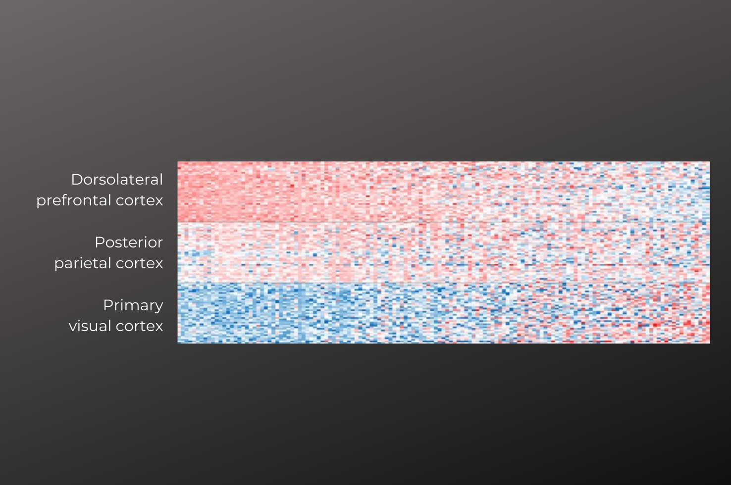
Findings demonstrated that the transcriptomes of layer 3 pyramidal neurons differ substantially across the cortical visual spatial working memory network and that these differences follow one of three patterns:
The transcriptome of layer 3 pyramidal neurons in the primary sensory cortex of this network differs greatly from those of layer 3 pyramidal neurons in the higher order association areas, posterior parietal and dorsolateral prefrontal cortices.
Other genes show gradients in expression along the rostral-to-caudal nodes of the visual spatial working memory hierarchy.
Some genes are specifically enriched in one region compared to the other two.
All these findings were highly conserved across individual subjects indicating that they reveal robust and reliable differences among layer 3 pyramidal neurons based on their regional location.
“These findings demonstrate that layer 3 pyramidal neurons are regionally-specialized to contribute in unique ways to information processing during working memory tasks and they provide the groundwork for examining how alterations in these neurons might contribute to illnesses, such as schizophrenia, that are characterized by impairments in working memory,” said Dr. Lewis, the study’s corresponding author.
Differential gene expression in layer 3 pyramidal neurons across 3 regions of the human cortical visual spatial working memory network
Enwright JF, Arion D, MacDonald WA, Elbakri R, Pan Y, Vyas G, Berndt A, Lewis DA.
Cerebral Cortex, 2022; https://doi.org/10.1093/cercor/bhac009
