Using Resting State fMRI to Study Everything but Neuronal Activity
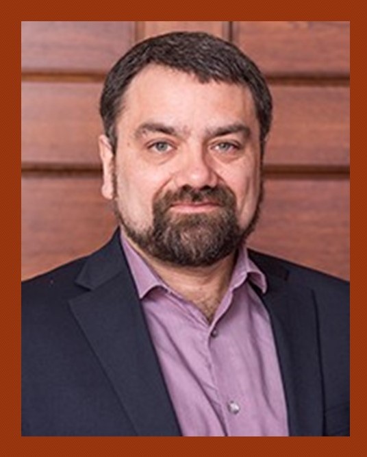
We hope you will join us for a Special Lecture on January 28, 2020 by Blaise Frederick, PhD.
 Dr. Frederick is an associate professor in psychiatry at Harvard Medical School and an associate biophysicist at McLean Hospital. He is also director of the Technical and Instrumentation Core at the McLean Imaging Center.Dr. Frederick received a BS in physics from Yale University and a PhD in biophysics from the University of California at Berkeley. His training is in MR physics and his PhD thesis is entitled “Three Dimensional Nuclear Magnetic Resonance Spectroscopic Imaging of Sodium Ions Using Stochastic Excitation and Oscillating Gradients.” Dr. Frederick is also the director of the McLean Imaging Center’s Opto-Magnetic Group, and his current research is focused on multimodal acquisition and processing for hemodynamic quantitation and physiological denoising of BOLD data and device development for clinical evaluation of peripheral vascular physiology.
fMRI is one of the most widely used techniques for studying human brain activity in vivo. The noninvasive nature of the method, in combination with very high spatial (and somewhat high temporal) resolution, wide availability, and good sensitivity led to its popularity in locating and quantifying neuronal activity. In recent years, resting state fMRI (rs-fMRI) has become increasingly common to examine networks of connectivity between brain regions in health and disease. But up to 50% of the signal variance in rs-fMRI is not due to neuronal activity. Long dismissed as simply “systemic physiological noise” and seen as a nuisance, the non-neuronal component of the fMRI signal carries rich information about cerebrovascular function and health. Dr. Blaise and his colleagues have developed and tested methods to extract regional blood flow information from the low frequency components of the rs-fMRI data without special acquisitions or additional measurements, and have evaluated these methods in moyamoya disease, stroke, and Alzhiemer’s disease, and in healthy comparison subjects to map vascular territories. More recently, the group has developed a method to extract high quality cardiac waveforms from the same datasets, and to map the pulsatile flow of blood throughout the brain, again using only resting state data with no additional measurements. This substantially increases the value of existing datasets, allowing detailed characterization of vascular function even if that was not considered in the design of the study, turning noise into signal. Finally, by parsing, modelling, and removing these signals, Dr. Blaise can effectively delete the majority of the non-neuronal components from rs-fMRI data (and most task data, according to our initial tests), removing two major confounds from accurate quantification of neuronal activation and connectivity.
Date and Time. Tuesday, January 28, 2020 from 12:00pm to 1:00pm
Location. Starzl Biomedical Science Tower, Room W1695.
For More Information. Please contact Yiming Wang via email at wangy40@upmc.edu.
